



Sumit Agrawal
Published Aug 31, 2026
New Synchrotron Imaging Findings: Implications for Individualized Cochlear Implantation
We have now long understood the extensive variability in cochlear size, shape, and morphology, and in recent years, research into this topic has increased. The study of cochlear length dates back to the early 19th century, but recent imaging research has revealed more insights into cochlear anatomy. With the advancement of clinical imaging and software, individual assessment is now possible as part of a clinical routine.
The Significance of Cochlear Variability
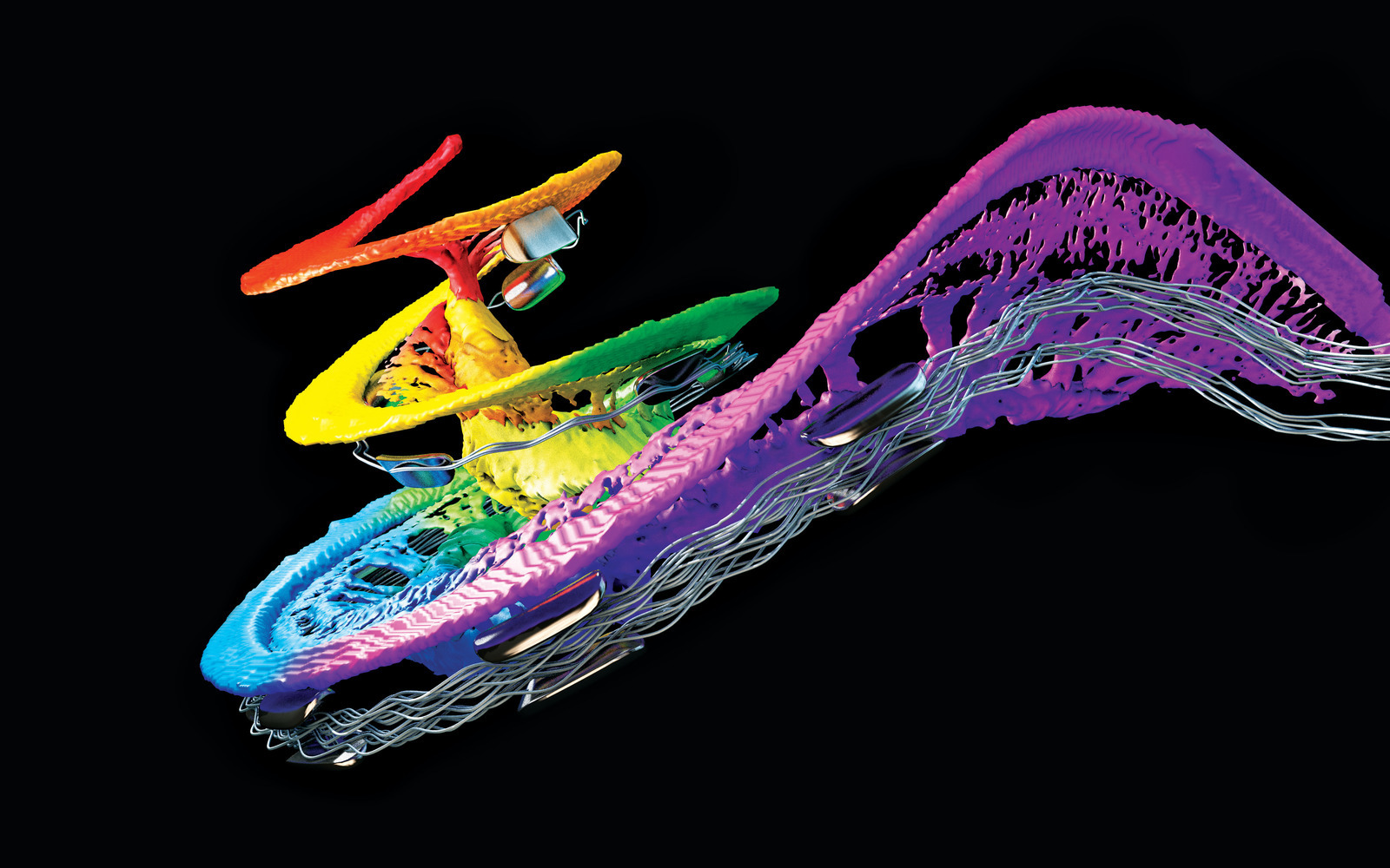
Cochlear length has historically been identified as a key parameter demonstrating considerable variability across individuals. Extensive variability has also been identified in other cochlear variables such as scala tympani size and volume.
This variability has significant implications for cochlear function and cochlear implantation. To understand individual factors such as cochlear length, tonotopic distribution, and scalar size, individual cochlear anatomic assessment is critical.
Incorporating individual anatomic assessment in clinical decisions makes it possible to preoperatively plan for optimal cochlear implant coverage while minimizing the risk of trauma. MED-EL strives to individualize cochlear implantation through different electrode array offerings, preoperative planning, and postoperative anatomy-based fitting.
Preoperatively, OTOPLAN allows for accurate and streamlined individualization in a clinical setting through visualization of cochlear anatomy and insertion predictions, enabling anatomy-based electrode selection. OTOPLAN also allows for postoperative image analysis and provides the measurements needed for anatomy-based fitting. In order to continue advancing models for individualization, research on the topic of cochlear variability is critical.
A Researcher and Surgeon’s Insights Into Cochlear Variability
Dr. Sumit Agrawal
Research in Cochlear Variability
Although there have been extensive studies into the variability of cochlear measures, such as cochlear duct length, these studies have typically had limitations in the modality or sample size of the data.
Histology was historically the only modality which allowed for accurate assessment and measurement of intracochlear anatomy, and multiple foundational studies demonstrated variability in cochlear anatomy using histology data.[1][2][3] However, due to the sample preparation and dissection required, the 3D anatomical structure is often not preserved in histology. Despite serving as an invaluable dataset for some cochlear anatomic analysis, it is not feasible to make certain measurements using these samples.
More recent imaging-based studies have provided 3D context for cochlear measurements. Computed tomography (CT) imaging has gained attention for its value in collecting cochlear measurements since it preserves 3D structures. Clinical and research CT imaging, such as micro-CT, have been used in numerous studies to measure cochlear length and dimensions.[4][5][6][7] The resolution and characteristics of these imaging modalities generally does not, however, contain adequate soft tissue contrast for ground truth intracochlear measurement.
In recent years, our Auditory Biophysics laboratory at Western University has used synchrotron imaging to successfully visualize intracochlear structures in intact cadaveric cochleae. This synchrotron imaging data has allowed for accurate soft tissue intracochlear measurements while preserving 3D morphology. Our previous publications have illustrated the use of synchrotron imaging for measuring cochlear length and scalar parameters accurately, but these studies have generally contained limited datasets, meaning they cannot capture the variability in cochlear size as extensively.[8][9][10][11]
The Latest Research: The Cochlear Morphometry Compendium
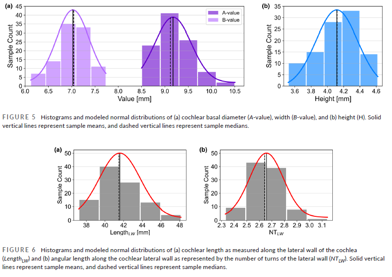
Our group has recently published a landmark reference measurement study titled “The cochlear morphometry compendium: High-resolution synchrotron measurements and normative refrence values.”[12] This study presents a large collection of reference cochlear measurements, derived from synchrotron imaging scans of 100 cadaveric cochleae.
This represents the first atlas of high-resolution measurements in a sample size this large and provides valuable data for the further development of individualized solutions. Reference measurements in the study include various cochlear duct lengths, tonotopic frequency distributions, cross-sectional scalar dimensions, scalar volume, and round window dimensions.
Source: Agrawal et al., 2026. https://doi.org/10.1111/joa.70175
Cochlear Duct Length and Tonotopic Frequency
Our synchrotron reference study analyzed complete cochlear length as well as proportional cochlear lengths at each angular depth in the synchrotron data collected from the 100 cochleae. Additionally, measurements such as the number of cochlear turns and the length of the basal hook region were reported. Complete cochlear duct length measurements (at the level of the organ of Corti) span from 30 mm to 40 mm in the reported values. This demonstrates significant variability and aligns with normative ranges reported in previous work. This highlights the value of various arrays to achieve desired cochlear coverage no matter the size of the cochlea.
Using Greenwood’s function, cochlear depths were related to tonotopic frequency. The significant variability in cochlear length results in variability in tonotopic frequency along the cochlea and highlights the need for individualization to achieve appropriate frequency coverage. This data also reinforces the value of apical insertions to achieve low frequency stimulation in the second turn of the cochlea.
Scalar Dimensions and Volume
Both scalar cross-sectional measurements and overall scalar volumes are reported in the study. Scalar volumes demonstrated significant variability, with the total scala tympani volume ranging from approximately 25 to 45 mm. Despite this variability, cross-sectional measurements revealed that even the smallest cochleae maintain sufficient area to accommodate modern flexible electrode arrays in the second turn.
Round Window Dimensions
In addition to expanding the dataset size for common clinical measures such as cochlear length and scalar dimensions, the synchrotron reference study established reference data for round window size. Considerable variability was observed in round window dimensions, with average long-axis and short-axis measurements of 2.20 ± 0.21 mm and 1.64 ± 0.18 mm, respectively. Round window shape and symmetry change drastically, but the average size confirms accommodation of arrays without requiring potentially traumatic round window extensions or cochleostomies. The variability, however, highlights the value of preoperative planning to understand what will be encountered during the procedure.
The expanded dataset collected and measurements made from synchrotron imaging make the extent of cochlear variability clearer than ever before. With image-based electrode selection along with anatomy-based fitting, surgeons can do justice to this cochlear variability by providing cochlear implants individualized to patients’ cochleae.
See the Data for Yourself
The expanded dataset collected and measurements made from synchrotron imaging make the extent of cochlear variability clearer than ever before. With image-based electrode selection along with anatomy-based fitting, surgeons can do justice to this cochlear variability by providing cochlear implants individualized to patients’ cochleae. Read the latest study for more details.
Download StudyReferences
-
[1]
Hardy, M. The Length of the Organ of Corti in Man. The American Journal of Anatomy 62, 291–311 (1938).
-
[2]
Stakhovskaya, O., Sridhar, D., Bonham, B. H., & Leake, P. A. (2007). Frequency map for the human cochlear spiral ganglion: Implications for cochlear implants. JARO – Journal of the Association for Research in Otolaryngology, 8, 220–233. https://doi.org/10.1007/s10162-007-0076-9
-
[3]
Úlehlová, L., Voldřich, L., & Janisch, R. (1987). Correlative study of sensory cell density and cochlear length in humans. Hearing Research, 28(2–3), 149–151. https://doi.org/10.1016/0378-5955(87)90045-1
-
[4]
Breitsprecher, T., Dhanasingh, A., Schulze, M., Kipp, M., Dakah, R. A., Oberhoffner, T., … Weiss, N. M. (2022). CT imaging-based approaches to cochlear duct length estimation—a human temporal bone study. European Radiology, 32, 1014–1023. https://doi.org/10.1007/s00330-021-08189-x
-
[5]
Escudé, B., James, C., Deguine, O., Cochard, N., Eter, E., & Fraysse, B. (2006). The size of the cochlea and predictions of insertion depth angles for cochlear implant electrodes. Audiology and Neurotology, 11, 27–33. https://doi.org/10.1159/000095611
-
[6]
Schurzig, D., Timm, M. E., Batsoulis, C., Salcher, R., Sieber, D., Jolly, C., … Zoka-Assadi, M. (2018). A Novel Method for Clinical Cochlear Duct Length Estimation toward Patient-Specific Cochlear Implant Selection. OTO Open, 2. https://doi.org/10.1177/2473974×18800238
-
[7]
Würfel, W., Lanfermann, H., Lenarz, T., & Majdani, O. (2014). Cochlear length determination using Cone Beam Computed Tomography in a clinical setting. Hearing Research, 316, 65–72. https://doi.org/10.1016/j.heares.2014.07.013
-
[8]
Helpard, L., Li, H., Rohani, S. A., Rask-Andersen, H., Ladak, H. M., & Agrawal, S. (2021). Three-Dimensional Modeling and Measurement of the Human Cochlear Hook Region: Considerations for Tonotopic Mapping. Otology & Neurotology, 42. https://doi.org/10.1097/mao.0000000000003065
-
[9]
Koch, R. W., Elfarnawany, M., Zhu, N., Ladak, H. M., & Agrawal, S. K. (2017). Evaluation of Cochlear Duct Length Computations Using Synchrotron Radiation Phase-Contrast Imaging. Otology and Neurotology, 38, e92–e99. https://doi.org/10.1097/mao.0000000000001410
-
[10]
Li, H., Helpard, L., Ekeroot, J., Rohani, S. A., Zhu, N., Rask-Andersen, H., … Agrawal, S. (2021). Three-dimensional tonotopic mapping of the human cochlea based on synchrotron radiation phase-contrast imaging. Scientific Reports, 11, 1–8. https://doi.org/10.1038/s41598-021-83225-w
-
[11]
Micuda, A., Li, H., Rask-Andersen, H., Ladak, H. M., & Agrawal, S. K. (2024). Morphologic Analysis of the Scala Tympani Using Synchrotron: Implications for Cochlear Implantation. Laryngoscope. https://doi.org/10.1002/lary.31263
-
[12]
Agrawal, S. K., Li, H., Spedaliere, C., Rask‐Andersen, H., & Ladak, H. M. (2026). The cochlear morphometry compendium: High‐resolution synchrotron measurements and normative reference values. Journal of Anatomy. https://doi.org/10.1111/joa.70175
References
Sumit Agrawal
Sumit Agrawal, MD, FRCS(C) is the Chairman and City-Wide Department Head of the Department of Otolaryngology - Head & Neck Surgery at Western University and London Health Sciences Centre in London, Ontario, Canada. He completed his fellowship in Neurotology & Skull Base Surgery and his clinical interests include cochlear implantation, vestibular surgery, and lateral skull base tumours. He is co-director of the Auditory Biophysics Laboratory and his current research involves middle-ear biomechanics, surgical simulation, imaging techniques, and artificial intelligence.



Was this article helpful?
Thanks for your feedback.
Sign up for newsletter below for more.
Thanks for your feedback.
Please leave your message below.
CTA Form Success Message
Send us a message
Field is required
John Doe
Field is required
name@mail.com
Field is required
What do you think?
The content on this website is for general informational purposes only and should not be taken as medical advice. Please contact your doctor or hearing specialist to learn what type of hearing solution is suitable for your specific needs. Not all products, features, or indications shown are approved in all countries.
Sumit Agrawal
Sumit Agrawal, MD, FRCS(C) is the Chairman and City-Wide Department Head of the Department of Otolaryngology - Head & Neck Surgery at Western University and London Health Sciences Centre in London, Ontario, Canada. He completed his fellowship in Neurotology & Skull Base Surgery and his clinical interests include cochlear implantation, vestibular surgery, and lateral skull base tumours. He is co-director of the Auditory Biophysics Laboratory and his current research involves middle-ear biomechanics, surgical simulation, imaging techniques, and artificial intelligence.