MED-EL
Published Sep 05, 2022 | Updated February 29, 2024
Anatomy-Based Fitting: A New Tool for Improving Place-Pitch Match
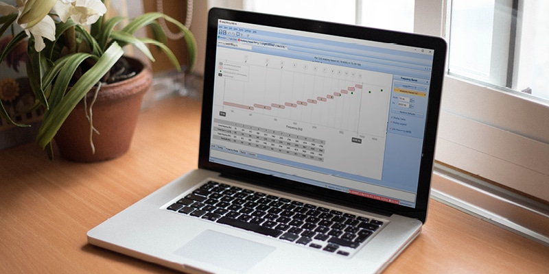
Clinicians are just starting to discover the great potential of anatomy-based fitting, an approach to cochlear implant frequency mapping that only MED-EL offers as part of our fitting software, MAESTRO, which is available to clinicians. It is the next step forward in providing cochlear implant recipients closer to natural hearing. As a new feature introduced with the release of MAESTRO 9.0, anatomy-based fitting (ABF) allows you to align the frequency map stimulated by the CI to the natural place-pitch frequency map in the cochlea as best as possible.
We all know how difficult it can be to find a comfortable pair of shoes that fit well. And when it comes to length, human cochleae have a wider range of variation than human feet do. That’s exactly why—preoperatively—it is not only important to find the best fit in terms of electrode array length. But—postoperatively—it is also important to do the fine tuning to reach the optimal hearing performance outcome.
Think of anatomy-based fitting as tying your shoes just right so they are neither too tight nor too loose on your feet. It’s also important that they fit your feet naturally without causing any discomfort after a long day. Of course, getting this feeling is only possible after you’ve put on a pair that is the right size.
Now, with more than a year’s field experience with anatomy-based fitting behind us, we’d like to share some of the real-world knowledge we have gained.
Does Anatomy-Based Fitting Work?
Here's the latest research on hearing outcomes and tonotopic mismatch.
Learn More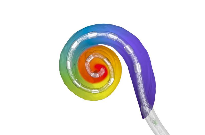
Anatomy-Based Fitting: The Working Principle
Our anatomy-based fitting tools help make it possible and easy to fine-tune the frequency map to be closer to the natural map for each individual cochlea—much like tying your shoes just right after you’re sure they are right size.
By importing post-op imaging data from OTOPLAN into MAESTRO, you can quickly assign patient-specific center frequencies based on the actual anatomical location of each electrode contact.* Based on the frequencies detected by OTOPLAN with post-op imaging, the software algorithm helps you to assign a more natural frequency distribution along the electrode array. This is now also possible with a post-operative x-ray.**
In simple terms, the main aim of anatomy-based fitting is to align each individualized fitting as closely to each individual recipient’s natural map as possible.
Insertion Depth, Insertion Angle & Tonotopic Frequency
The modifications that are made by anatomy-based fitting to the standard frequency bands depend on how far an electrode has been inserted into the cochlea.
The insertion depth (in mm), the insertion angle (in degrees) and the covered frequencies (in Hz) are all interrelated. Practically speaking, in normal cochleae, angle and frequency will always have a relatively fixed relationship. That means that, on average, 630° will be approximately at 180 Hz (Organ of Corti, OC). This is extremely important because if we know the angular location of the electrode contact, we can estimate the tonotopic frequency.
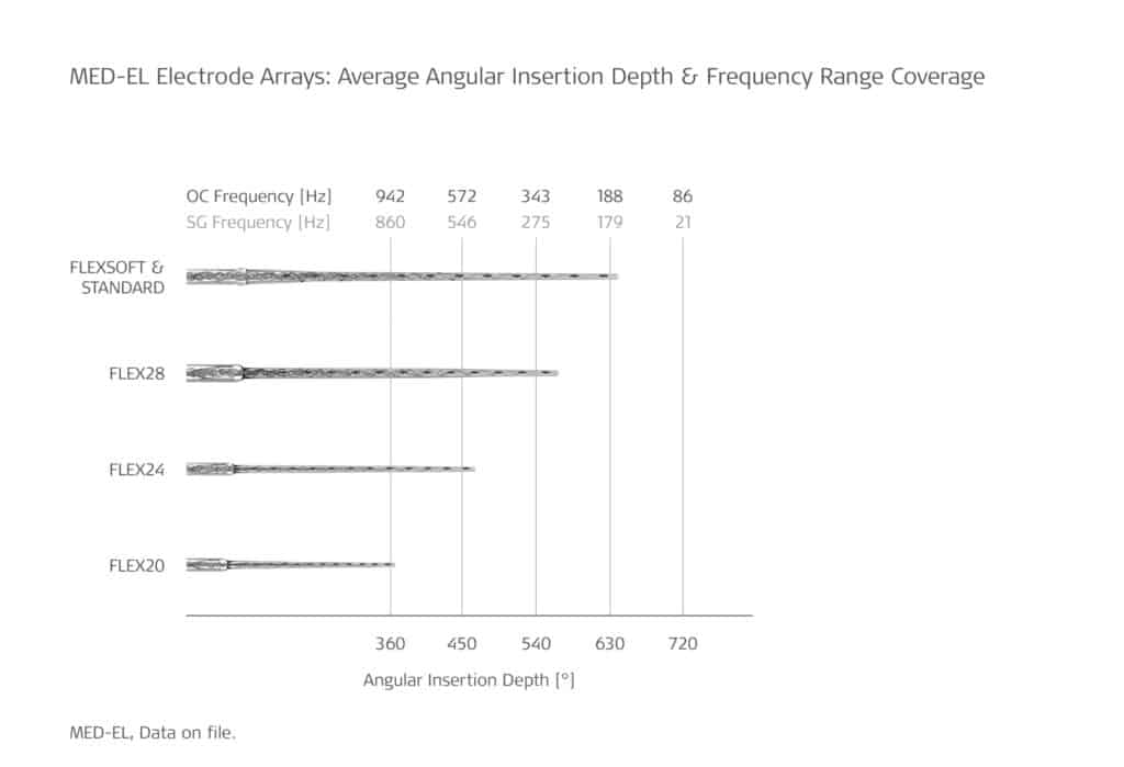
In Figure 1, you can see an analysis of insertion depth angles and frequency range covered by the first apical contact (E1) for different MED-EL electrodes. Note: The renderings shown here are not accurate depictions of the actual sizes of the cochlear implant electrode arrays. The FLEX34 electrode array is not included because it was not yet introduced.
The reason why MED-EL’s electrodes, measured in millimeters, come in different lengths is that cochleae come in different sizes. For instance, the same FLEX28 electrode array might achieve deep insertion (>630°) in a smaller cochlea, but shallower insertion in a larger cochlea (540° or less). Naturally, longer electrodes can reach further into the cochlea and potentially provide a better place-pitch match. Canfarotta, M. W., Dillon, M. T., Buss, E., Pillsbury, H. C., Brown, K. D., & O’Connell, B. P. (2020). Frequency-to-place mismatch: Characterizing variability and the influence on speech perception outcomes in cochlear implant recipients. Ear & Hearing, 41(5), 1349–1361. https://journals.lww.com/ear-hearing/Fulltext/2020/09000/Frequency_to_Place_Mismatch__Characterizing.27.aspx[1]Landsberger, D.M., Vermeire, K., Claes, A., Van Rompaey, V., & Van de Heyning, P. (2016). Qualities of single electrode stimulation as a function of rate and place of stimulation with a cochlear implant. Ear & Hearing., 37(3), 149–159. https://www.ncbi.nlm.nih.gov/pmc/articles/PMC4844766/[2]Li, H., Schart-Moren, N., Rohani, S., A., Ladak, H., M., Rask-Andersen, A., & Agrawal, S. (2020). Synchrotron Radiation-Based Reconstruction of the Human Spiral Ganglion: Implications for Cochlear Implantation. Ear Hear. 41(1). https://journals.lww.com/ear-hearing/Fulltext/2020/01000/Synchrotron_Radiation_Based_Reconstruction_of_the.18.aspx[3]
In general, MED-EL recommends aiming for complete cochlear coverage, achieved with insertion depths between 540° (1.5 turns) and 720° (2 turns), where insertion depths above 630° (1.75-2 turns) will potentially provide a more accurate and balanced anatomy-based fitting map. This means that MED-EL recommends aiming for a center frequency of electrode 1 (E1) between 340 Hz and 85 Hz.
This makes preoperative planning even more important in cases where post-operative ABF is desired. OTOPLAN can help with the planning process and in choosing a suitable electrode to achieve complete cochlear coverage. This enables the surgeon to have an influence on the postoperative fitting and potential outcome of the patient.
Note: In the sections below, the consequences that anatomy-based fitting has on the filter bands are discussed as a function of E1 frequency, which primarily depends on electrode insertion depth. All frequencies are at the level of Organ of Corti (OC) as displayed in the latest version of OTOPLAN.
Average Insertion (630°, approx. 185 Hz)
The standard frequency bands have been designed to correspond well with natural tonotopicity for E1 at approximately 185 Hz (equivalent to an insertion angle of 630°). This can typically be achieved with STANDARD, FLEXSOFT, and – in smaller cochleae – FLEX28 electrode arrays. In the example shown in Figure 2, notice that there is a small degree of variation between the standard frequency bands (illustrated in grey) and ABF (illustrated in color) for an E1 frequency of about 185 Hz. This is ideal because stimulated electrode frequencies match natural tonotopic place across most of the array, and the frequencies are well-distributed across each electrode channel. This allows the work to be “shared” across the whole array without too many frequencies sharing one contact.
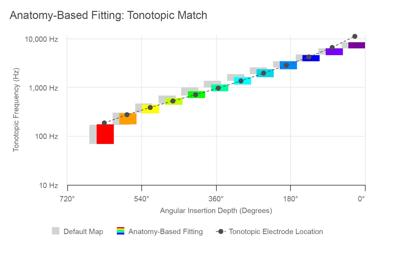
Figure 2. Default and anatomy-based filter bands for an average insertion with a STANDARD or FLEXSOFT electrode.
Deep Insertion: Complete Cochlear Coverage (720°, approx. 100 Hz)
FLEXSOFT and STANDARD electrode arrays reach deeper into the cochlea, thus more reliably achieving Complete Cochlear Coverage in most cochleae. This means they reach an E1 frequency of 100 Hz or lower (corresponding to an insertion angle of up to 720° or more). As indicated in Figure 3, with ABF, in this case some frequencies are shifted basally when compared to the standard filter bands. Here, the adjustment made by ABF is more significant compared to an average insertion. However, the frequency bands are well-balanced across the cochlea and the array so this can be considered an optimal outcome.
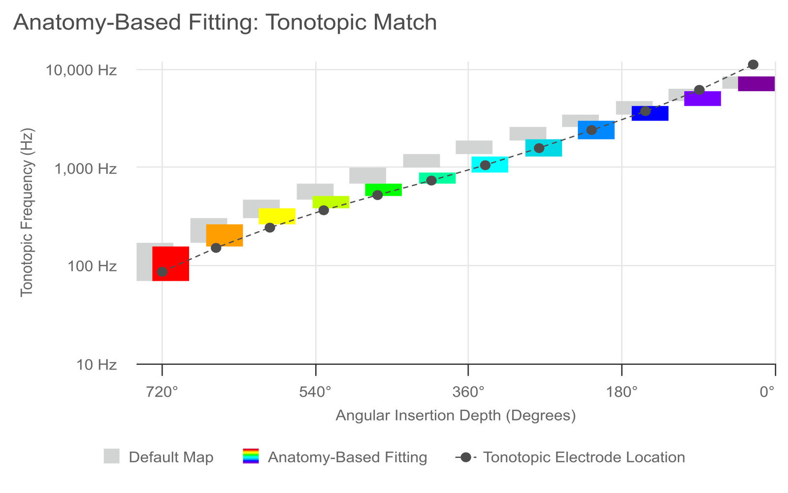
Figure 3. Default and anatomy-based fitting filter bands for a deep insertion with a STANDARD or FLEXSOFT electrode.
Insertion of 1.5 Turns (540°, approx. 340 Hz)
There is a wide range in the cochlear duct length between different cochleae. In smaller cochleae, a FLEX28 may provide sufficient coverage. However, in the average cochlea, FLEX28 electrode arrays mostly don’t reach far enough into the cochlea to provide true complete cochlear coverage. Instead, it often results in E1 frequencies of approximately 340 Hz (corresponding to 1.5 turns, or 540°).
With these insertions, ABF results in a moderate apical shift of bandpass frequencies. This means that a somewhat wider range of frequencies will be compressed onto the apical channels in order to allow tonotopic electrode frequencies between 950 Hz and 3 kHz, as this frequency range is a priority for speech understanding.
Shallow Insertion (<540°, <340 Hz)
The FLEX26 and FLEX24 array usually achieve insertions of less than 340 Hz (less than 1.5 turns). With these insertions, a significant apical shift of bandpass frequencies results. This means that a considerably wide range of frequencies will be compressed into the apical channels. For ABF to be utilized, at least 2 contacts must be below 950 Hz and at least 2 contacts must be between 950 Hz and 3 kHz. For example, ABF is usually not available in MAESTRO with insertions typically achieved with the FLEX20 electrode array (930 Hz, i.e. <1 turn) or even shallower insertions.
Anatomy-Based Fitting & Coding Strategies
ABF should preferably be used in conjunction with FineHearing coding strategies, i.e. FSP, FS4 or FS4-p. These strategies can extend further into the low frequencies by default. More information about FineHearing can be found here.
Anatomy-Based Fitting: Another Step Towards Optimized Hearing Outcomes
Anatomy-based fitting is a powerful tool that can be used to further optimize hearing outcomes. While research is still preliminary and a larger number of cases need to be observed before reaching more definitive conclusions, we can already recommend the following:
- The first step is making sure an electrode of the right length has been implanted and OTOPLAN can help with this. The deeper the electrode array insertion and the more cochlear coverage is achieved, the better the outcomes with anatomy-based fitting tend to be. As a result, FLEXSOFT and STANDARD arrays are best suited for anatomy-based fitting, and the FLEX28 array may also be considered a suitable option in some cases. In contrast, experience so far has shown that ABF in combination with shallow electrode insertion produces a rather ‘bassy’, dull or low-pitched sound perception.
- The second is that anatomy-based fitting requires a postoperative CT scan or plain x-ray, which may not yet be included in the clinical routine.** By knowing the potential added benefits of anatomy-based fitting, the time and resources invested for the post-op scan may be well worth it. Note that the 2024 release of OTOPLAN allows for anatomy-based fitting with a postoperative x-ray.
- The third is for clinicians to leverage post-op CT or plain x-ray imaging and incorporate anatomy-based fitting into their practice and post-operative fitting workflows with the MAESTRO fitting software.
By taking these steps, hearing outcomes can be further optimized for recipients. Having shoes that are the right size is one step towards all-day comfort when walking. But doesn’t it also make sense to make sure our shoes are properly tied to ensure the best possible fit? Anatomy-based fitting is the next step towards reaching optimal hearing performance outcomes for each individual cochlear implant recipient.
Hearing Insights in Your Inbox
Get the latest research and resources to help people with every kind of hearing loss. Subscribe to the MED-EL Professionals Blog now.
Sign Me Up!References
-
[1]
Canfarotta, M. W., Dillon, M. T., Buss, E., Pillsbury, H. C., Brown, K. D., & O’Connell, B. P. (2020). Frequency-to-place mismatch: Characterizing variability and the influence on speech perception outcomes in cochlear implant recipients. Ear & Hearing, 41(5), 1349–1361. https://journals.lww.com/ear-hearing/Fulltext/2020/09000/Frequency_to_Place_Mismatch__Characterizing.27.aspx
-
[2]
Landsberger, D.M., Vermeire, K., Claes, A., Van Rompaey, V., & Van de Heyning, P. (2016). Qualities of single electrode stimulation as a function of rate and place of stimulation with a cochlear implant. Ear & Hearing., 37(3), 149–159. https://www.ncbi.nlm.nih.gov/pmc/articles/PMC4844766/
-
[3]
Li, H., Schart-Moren, N., Rohani, S., A., Ladak, H., M., Rask-Andersen, A., & Agrawal, S. (2020). Synchrotron Radiation-Based Reconstruction of the Human Spiral Ganglion: Implications for Cochlear Implantation. Ear Hear. 41(1). https://journals.lww.com/ear-hearing/Fulltext/2020/01000/Synchrotron_Radiation_Based_Reconstruction_of_the.18.aspx
References
MED-EL
Was this article helpful?
Thanks for your feedback.
Sign up for newsletter below for more.
Thanks for your feedback.
Please leave your message below.
CTA Form Success Message
Send us a message
Field is required
John Doe
Field is required
name@mail.com
Field is required
What do you think?
The content on this website is for general informational purposes only and should not be taken as medical advice. Please contact your doctor or hearing specialist to learn what type of hearing solution is suitable for your specific needs. Not all products, features, or indications shown are approved in all countries.
MED-EL


