Search
Search
Menu
Close
MED-EL
Published Sep 05, 2018

The MED-EL Surgical Video Library offers complete surgical case studies from leading ENT surgeons. Created in cooperation with ARRI, these high-resolution videos capture precise movements and detailed structures with incredible clarity. Access is free and the easy-to-use library is optimized for desktop or mobile viewing.
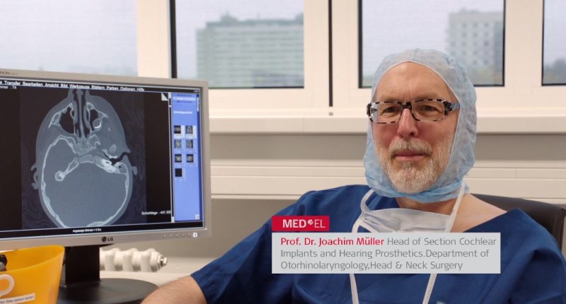
In this step-by-step surgical case study, Prof. Dr. Joachim Müller guides us through his techniques for implanting the SYNCHRONY Cochlear Implant. Prof. Müller is the Head of the Cochlear Implants and Hearing Prosthetics Section, Department of Otorhinolaryngology, Head & Neck Surgery at the University of Munich.
In this case, Prof. Müller’s patient is a 2-year-old female with profound bilateral sensorineural hearing loss. This is the second surgery of a sequential bilateral implantation; the patient had already been implanted 9 months earlier with a cochlear implant in her contralateral (right) ear.
In this high-definition surgical video, Prof. Müller utilizes a SYNCHRONY Cochlear Implant with Standard electrode array for the patient’s left ear. The 31.5 mm Standard array enables apical stimulation while allowing for preservation of delicate cochlear structures. Prof. Müller also utilizes the MED-EL Fixation Clip to further secure the placement of the electrode array.
Watch now: Prof. Joachim Müller demonstrates his surgical techniques for implanting the SYNCHRONY Cochlear Implant and Standard electrode array. [24 minutes]
What to watch for:
Want to see more videos from Prof. Müller? Check out his surgical techniques for the BONEBRIDGE Active Bone Conduction Implant.
Have a question about surgical techniques for SYNCHRONY? Let us know with our contact form or leave a comment below.
*Not all products, features, and indications shown are available in all areas. Please contact your local MED-EL representative for more information.
MED-EL
Was this article helpful?
Thanks for your feedback.
Sign up for newsletter below for more.
Thanks for your feedback.
Please leave your message below.
CTA Form Success Message
Send us a message
Field is required
John Doe
Field is required
name@mail.com
Field is required
What do you think?
The content on this website is for general informational purposes only and should not be taken as medical advice. Please contact your doctor or hearing specialist to learn what type of hearing solution is suitable for your specific needs. Not all products, features, or indications shown are approved in all countries.
MED-EL
Get the latest research and resources to help people with every kind of hearing loss. Subscribe to the MED-EL Professionals Blog now.
Registration was successful
We’re the world’s leading hearing implant company, on a mission to help people with hearing loss experience the joy of sound.
Find your local MED-EL team
The content on this website is for general informational purposes only and should not be taken as medical advice. Please contact your doctor or hearing specialist to learn what type of hearing solution is suitable for your specific needs. Not all products, features, or indications shown are approved in all countries.


