Join Clinicians Worldwide
Evidence-based insights to enhance hearing care - twice a month
Subscribe Now Evidence-based insights to enhance hearing care - twice a month
Subscribe NowMED-EL
Published Dec 06, 2018

The MED-EL Surgical Video Library offers complete surgical case studies from leading ENT surgeons. Created in cooperation with ARRI, these high-resolution videos capture precise movements and detailed structures with incredible clarity. Access is free and the easy-to-use library is optimized for desktop or mobile viewing.
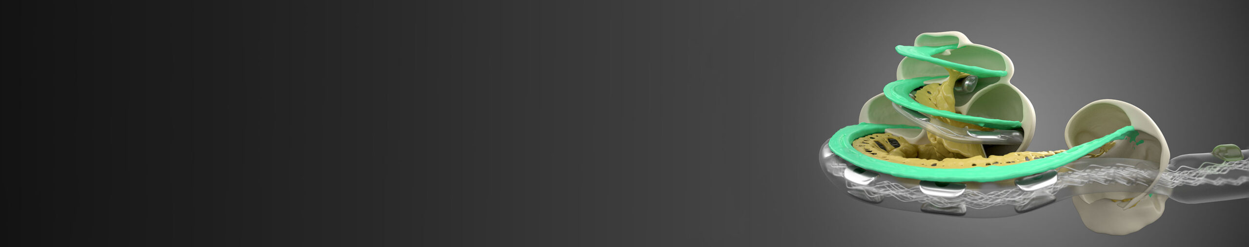
In today’s HD surgical case study, Dr. Harold Pillsbury guides us through his technique with the MED-EL CONCERT cochlear implant. Dr. Pillsbury is the Chair of the Department of Otolaryngology/Head and Neck Surgery at University of Carolina School of Medicine.
This patient is a 64-year-old woman with progressive bilateral hearing loss, and this is her second cochlear implant. The pre-operative audiogram shows minimal-to-no residual hearing. Despite multiple comorbidities, (hypertension, heart attack, Crohn’s disease), there are no complications in this case.
Dr. Pillsbury narrates the surgical video and provides valuable insight into his surgical techniques. He also explains why you cannot use monopolar cautery on a patient with a cochlear implant. In this case, the patient received a MED-EL CONCERT implant with a 31.5 mm Standard electrode array. However, these techniques are also applicable for SYNCHRONY.
One-month post-operative audiogram shows excellent results, with HINT scores going from 0% to 92% for the implanted ear.
Watch now: Dr. Harold Pillsbury guides us through his technique for implanting a MED-EL cochlear implant with 31.5 mm Standard electrode array (15 min).
What to watch for:
*Not all products, indications, and features shown are available in all areas. Please contact your local MED-EL representative for more information.
MED-EL
Was this article helpful?
Thanks for your feedback.
Sign up for newsletter below for more.
Thanks for your feedback.
Please leave your message below.
CTA Form Success Message
Send us a message
Field is required
John Doe
Field is required
name@mail.com
Field is required
What do you think?
The content on this website is for general informational purposes only and should not be taken as medical advice. Please contact your doctor or hearing specialist to learn what type of hearing solution is suitable for your specific needs. Not all products, features, or indications shown are approved in all countries.
MED-EL
Get the latest research and resources to help people with every kind of hearing loss. Subscribe to the MED-EL Professionals Blog now.
Registration was successful
We’re the world’s leading hearing implant company, on a mission to help people with hearing loss experience the joy of sound.
Find your local MED-EL team
The content on this website is for general informational purposes only and should not be taken as medical advice. Please contact your doctor or hearing specialist to learn what type of hearing solution is suitable for your specific needs. Not all products, features, or indications shown are approved in all countries.