
Join Clinicians Worldwide
Evidence-based insights to enhance hearing care - twice a month
Subscribe Now Evidence-based insights to enhance hearing care - twice a month
Subscribe NowMED-EL
Published Jul 26, 2023
A new user interface, fully automatic 3D reconstruction and measurement of inner ear structures, simultaneous preoperative visualizations of various electrode arrays in the cochlea, support for placement planning of cochlear and bone conduction implants, and extensive postoperative analysis tools—those are just a few of the innovative features in the newest version of OTOPLAN. This software solution makes it possible to individualize hearing implant surgery—from planning the operation to the audiological fitting—like never before.

An absolute highlight of the newest OTOPLAN software is automatic reconstruction of the entire inner ear in less than two minutes. Using the preoperative imaging, OTOPLAN creates a detailed model of the inner ear structures automatically: cochlea, round window membrane, bony overhang, semicircular canals, and internal auditory canal. Zoom in on the 3D reconstruction and rotate it in any direction with the 360° view.

If needed, the 3D model can be exported as an STL file and used to make a 3D print of the inner ear. OTOPLAN can process all standard MRI and CT formats and use them as a basis for visualizations: MDCT, CBCT as well as DVT. The images can be imported as DICOM files, and OTOPLAN is also compatible with PACS and a variety of external data storage devices, such as USB drives, DVDs, and CDs.
There is an additional brand-new feature connected to 3D reconstruction: fully automatic measurement of the cochlea, including cochlear duct length. A variety of tools for manual measurement (linear, polygonal, spline, freeform, angle measurement, shortest distance between 3D structures) are also available.
After creating a 3D reconstruction of the anatomy of the temporal bone and placing a target point, the 3D models can be rotated freely around this point to find the optimal trajectory for surgical access to the cochlea. Additionally, OTOPLAN can automatically calculate the distances between the planned trajectory and critical anatomical structures.
In addition to bony and neural structures, the newest version of OTOPLAN can automatically create a 3D reconstruction of the skin, map skin flap thickness based on a custom threshold, and create a color-coded anatomical heat map. This data can be used to plan the position of the implant housing and audio processor preoperatively. For the most asthetically appealing result in the case of bilateral implantation, OTOPLAN can mirror the position of the audio processor to the contralateral side.

There is another great improvement in preoperative implant placement planning: It’s also available for the BCI 602—not just cochlear implants.
In addition to skin thickness mapping, bone thickness mapping is also available for BONEBRIDGE implantation. And it is even possible to fit the skull with the BCI 602 implant virtually. The colors on the heat map show which locations would be most suitable for implant placement based on the bone thickness map. The corresponding 3D model can be displayed with the implant placed in the bone virtually, or it can be shown without the implant but with the recess in the bone that serves as the implant bed.

Another new feature that makes preoperatively estimating the position of the electrode easier than ever before is the so-called 3D Audiogram. It combines anatomical data (3D reconstruction) and audiological data (audiogram). Every 3D audiogram mirrors the individual anatomical and audiometric conditions and illustrates them with the help of intuitive color coding. In the end, users receive a representation of the patient’s cochlea in which the level of color saturation makes the individually-measured hearing loss visible at a glance.

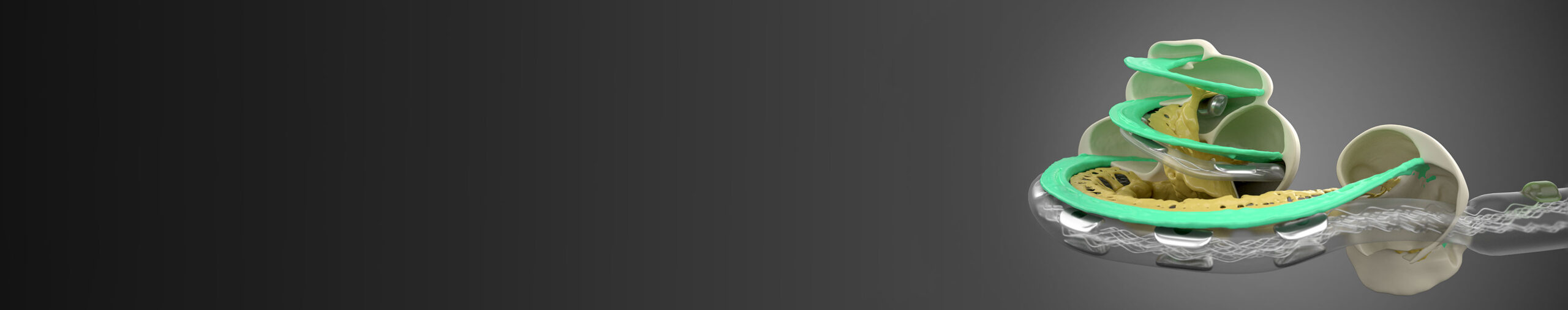
In the last version of OTOPLAN, it was possible to see preoperatively how an electrode array would fit inside the cochlea and display the corresponding frequency and insertion angle for each electrode contact. Now the visualization of two different electrode lengths at the same time is possible. OTOPLAN can show two electrode arrays on top of each other in the cochlea and compare the position of the contacts, including angular insertion depth and tonotopic center frequency. In addition, the brand-new FLEX34 electrode array is already in the newest version of OTOPLAN.

"Preoperative visualization of various electrodes in the cochlea makes it much easier to select the correct electrode array, especially in cases of partial hearing loss. It is particularly useful that it is possible to export the exact position of the electrode contacts after the operation. This data can be used after implant activation for audiological fine tuning."
Philipp Schörg, BSc, MSc
Deputy Director of Logopedics and Audiology, University Hospital, St. Pölten
OTOPLAN offers a wide range of possibilities to fuse different kinds of pre- and postoperative images (MRI, CT) together. Potential uses include image fusion of CT and MRI scans for an overview of all relevant types of tissue—bones, nerves, and soft tissue—or the fusion of a postoperative CT image with preoperative CT measurements of the cochlea. That has the advantage that audiologists don’t need to take measurements again after implantation.
Automatic fusion of up to four different images is possible with OTOPLAN’s image fusion.
“The automatic inner ear 3D reconstruction and cochlear measurement features make the electrode selection process more efficient.The image fusion feature gives us more anatomical information and better planning possibilities by automatically overlaying the images from different modalities.”
Dr. Lukas Vargas, MSc, PhD
ENT Surgeon, Comenius University Clinic, Bratislava
Automatic detection of the exact position of each electrode contact in the cochlea was already standard in the previous version of OTOPLAN, but now this frequency and localization data—which is necessary for anatomy-based fitting (ABF)—can be exported with the new version of OTOPLAN in seconds, saved externally, and/or uploaded into the MAESTRO fitting software (version 9 or later).
OTOPLAN can detect the electrode contacts automatically on the CT scan, as well as the implant housing and the electrode lead. With the post-op analysis, it is possible to perform a quality check of the lead management status. The detected position of the implant housing can be used to place the second implant as symmetrically as possible on the contralateral side.

Another new feature is screen recording. Starting and stopping a screen recording at any time is possible in the new version of OTOPLAN. Workflow screen recordings can be useful for training, documentation, and quality assurance. Additionally, there is the option to record audio commentary simultaneously with a microphone.
Would you like to use the newest version of OTOPLAN, need training with the software, or simply have questions about OTOPLAN? Don’t hesitate to get in touch with your local MED-EL team or representative. We’re happy to help you with all of your questions.
OTOPLAN is downward compatible—after upgrading to the newest version, you can immediately continue working on all cases from the previous version. The new version of OTOPLAN is delivered without hardware and works on Windows laptops, tablets, and PCs.
Subscribe now to the MED-EL blog for professionals and receive regular updates from MED-EL directly to your inbox.
References
MED-EL
Was this article helpful?
Thanks for your feedback.
Sign up for newsletter below for more.
Thanks for your feedback.
Please leave your message below.
CTA Form Success Message
Send us a message
Field is required
John Doe
Field is required
name@mail.com
Field is required
What do you think?
The content on this website is for general informational purposes only and should not be taken as medical advice. Please contact your doctor or hearing specialist to learn what type of hearing solution is suitable for your specific needs. Not all products, features, or indications shown are approved in all countries.
Siegfried dalpetz
July 27, 2023
I receiveda cocholear implant in my life year to years ago in London Ontario Canada,I have a cocholear inplant commit up i’m September 11 on my right ear would they be using the latest technology and equipment for the implant? My previous implant was done on my left ear. Please let me know. Siegfried f. Daloetz
MED-EL
July 28, 2023
Hi Siegfried, thanks for your comment. Your medical professional is best equipped to help you in this case, so we recommend reaching out to them as every surgery depends on the individual requirements. Kind regards, Gordana
MED-EL
Get the latest research and resources to help people with every kind of hearing loss. Subscribe to the MED-EL Professionals Blog now.
Registration was successful
We’re the world’s leading hearing implant company, on a mission to help people with hearing loss experience the joy of sound.
Find your local MED-EL team
The content on this website is for general informational purposes only and should not be taken as medical advice. Please contact your doctor or hearing specialist to learn what type of hearing solution is suitable for your specific needs. Not all products, features, or indications shown are approved in all countries.


Conversation
1 Comment