

Join Clinicians Worldwide
Evidence-based insights to enhance hearing care—twice a month
Subscribe Now Evidence-based insights to enhance hearing care—twice a month
Subscribe Now
Published Jan 17, 2024
With endoscopic ear surgery, surgeons can get a wider field of view when implanting passive middle ear prostheses for ossicular reconstruction and stapes surgery. Learn what tips and tricks Prof. Dr. med. Lukas Anschütz has for using an endoscope while implanting tympanoplasty and stapes prostheses from MED-EL.

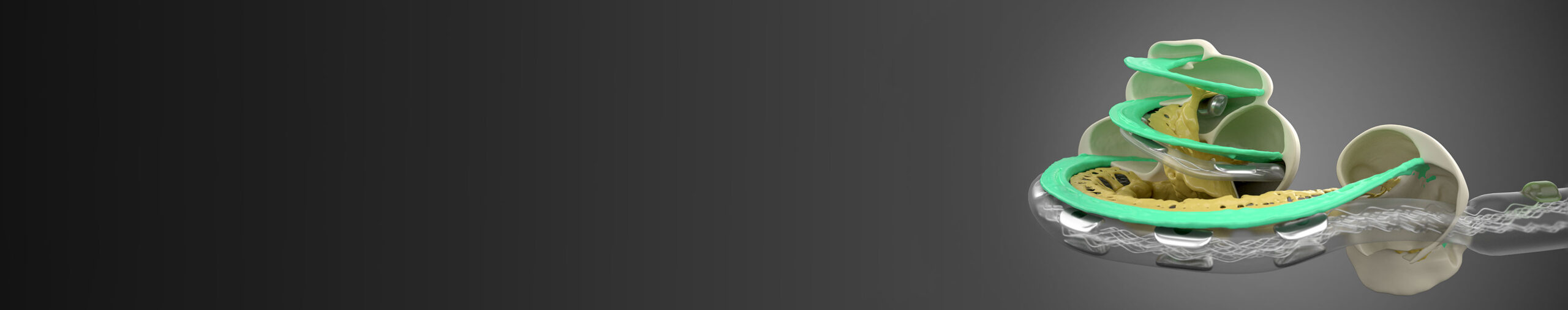
Passive middle ear implants are the most recent addition to MED-EL’s portfolio of hearing solutions. They serve as replacements for the tiny bones of the middle ear’s ossicular chain. MED-EL offers both partial and total tympanoplasty prostheses as well as stapesplasty prostheses for a total of 11 designs. They are made of biocompatible, durable titanium and allow for MRI scans at 1.5, 3.0, and 7.0 Tesla.
Designed with simple surgery in mind, passive middle ear implants provide an optimal interoperative view and excellent adaptability. Surgeons can adjust the length of the prostheses without any pre-set increments, and adjustment requires no special tools. Sizers are included in the kit the prosthesis kits.
In this SurgeryONLINE, Prof. Dr. med. Lukas Anschütz from the ENT Surgery Department at the University Hospital of Bern presents some principles of endoscopic ear surgery and demonstrates how he uses the endoscope to perform ossicular reconstruction and stapes surgery.

Performing endoscopic surgery has a variety of advantages over the typical approach using a microscope. It provides a wide field of view, and this ability to see—and show—more is particularly good for teaching. An endoscope magnifies the structures of the ear’s anatomy and provides the surgeon with the ability to look around the corner. Additionally, this type of surgery increases preservation of the mastoid and the external ear canal. At 30-50% power, there is enough light to see without heating up the middle ear too much.
Although this way of performing surgery is less invasive, there is less room to work. Surgery must therefore be performed using only one hand, which comes with a learning curve. Right-handed surgeons may want to begin with a surgery on the left side and vice versa. Less space also means that suction to control bleeding is limited. A 3mm endoscope means limited workspace in the canal, which does not provide the flexibility needed when there is disease inside the mastoid. Such situations require a combined approach.

Dr. Anschütz shares four different cases using the mXACT partial, the mCLIP ARC partial, and the mAXIS stapes prostheses.
This patient was in general good health but was referred to Prof. Anschütz due to a suspected cholesteatoma. Based on the pre-operative CT scan, the cholesteatoma appeared to be small enough for endoscopic surgery.
Dr. Anschütz uses a variety of techniques for hemostasis: adrenaline and lidocaine injections, monopolar cautery at the lowest intensity (to prevent damage to the facial nerve) before cutting the flap, as well as neuro-patties soaked in pure adrenaline. After elevating the flap, Prof. Anschütz dissects the cholesteatoma from the rest of the flap. Then he can pull the flap anteriorly and inferiorly to get it out of the way for the further dissection. Then he carefully removes a little bit of bone with a piezoelectric device. An advantage of this method is that it does not cut soft tissue. He does this under water to have a good view of the bone removal.
Then he cuts the incudostapedial joint with a round knife and then removes the incus and the head of the hammer to be able to further dissect and remove the cholesteatoma. With a 45 degree endoscope he is able to look into the antrum where he detects one last small piece of cholesteatoma and then removes it with suction.
With narrow-band imaging (digital image enhancement technology), he can make sure he has not overlooked any cholesteatoma. With the endoscope, he also sees that the tensor fold is completely closed which may have been the reason for the cholesteatoma in the first place.
He opens the tensor fold to improve ventilation during the healing process, places the mCLIP ARC partial prosthesis onto the stapes, and then covers the prosthesis headplate with a piece of cartilage. The mCLIP ARC has a micro-ball joint that connects the headplate to the shaft. Although it might seem challenging to place this prosthesis with the endoscope, it is actually easily feasible.
The second case is very similar to the first one, but on the CT scan, it is clear to see that the cholesteatoma is larger. Similar methods to those used in the first case are utilized to reduce bleeding, and Prof. Anschütz uses a piezoelectric device to remove bone. Then he dissects the incudostapedial joint, removes the remaining part of the incus, and cuts off and removes the head of the hammer. The chorda tympani must also be cut for safety reasons since it is inside the cholesteatoma. Prof. Anschütz recommends using a Thomassin dissector to clean the cholesteatoma away.
After removing all of the cholesteatoma that extended into the mastoid, he uses a 45 degree endoscope and digital image enhancement to check for remaining cholesteatoma.
The method for placing the mCLIP ARC is slightly different in this case: Prof. Anschütz uses suction to place the mCLIP ARC onto the stapes and then pushes it down gently. The steps that follow are similar to the first case, and the dressing of the external auditory canal using silk (which is not sticky and will hardy be felt when it is removed) is also shown.
Although the patient was initially happy with the results of the first surgery in 2015, a revision surgery is necessary since antibiotics for otorrhea were not effective. The pre-op CT scan shows that the middle ear and mastoid are completely full of granulation tissue.
Most surgeons would say a mastoidectomy is necessary, but Prof. Anschütz shows that a surgeon with enough experience can successfully perform an endoscopic surgery under these conditions. The ear is very inflamed and contains scar tissue from the previous implantation.
Prof. Anschütz removes the incus interposition from the previous surgery as well as the granulation tissue from the stapes. Then he places the mXACT partial prosthesis onto the stapes and places a piece of cartilage on top of the prosthesis before he closes the tympanomeatal flap.
This patient had otosclerosis of the stapes, and although a CT scan is not typically done to check this, it can show hints of otosclerosis and be used to check for and exclude other pathologies. As in Case 1, monopolar cautery is used before cutting the tympanomeatal flap. Compared to the cholesteatoma cases shown before, the flap is a little smaller because less bone needs to be removed.
Prof. Anschütz checks the mobility of the ossicles and confirms that the stapes is fixed. He cuts the incudostapedial joint and then uses a diode laser to cut through the stapedial tendon and posterior crus. (A microdrill or crura scissors can also be used.) Then he breaks away the anterior crus and removes the stapes superstructure. With a 0.6 mm diamond drill, he makes a hole in the footplate.
Then he measures the distance from the stapes footplate to the lateral side of the incus to figure out the correct prosthesis length. Using forceps and a hook for positioning, he then places the mAXIS stapes prosthesis. And finally, he closes the implant around the long process of the incus.
Endoscopes bring innovation into ear surgery and provide excellent visualization while making minimally invasive procedures possible—they result in practically no scars. Endoscopic ossiculoplasty using a titanium prosthesis is a straightforward procedure. It is important for surgeons to consider their learning curve and choose their first cases wisely. For more advanced cases, a combined approach can be taken. To learn more, take a look at this meta-analysis on the endoscopic approach to middle ear surgery and this article about mastoid preservation.
The MED-EL Surgical Video Library offers complete surgical case studies from leading ENT surgeons with step-by-step narration. These high-definition videos were created in collaboration with ARRI and show precise movements and detailed structures with incredible clarity. With your free myMED-EL professionals account, you get unlimited access to the MED-EL Surgical Video Library.
References
Was this article helpful?
Thanks for your feedback.
Sign up for newsletter below for more.
Thanks for your feedback.
Please leave your message below.
CTA Form Success Message
Send us a message
Field is required
John Doe
Field is required
name@mail.com
Field is required
What do you think?
The content on this website is for general informational purposes only and should not be taken as medical advice. Please contact your doctor or hearing specialist to learn what type of hearing solution is suitable for your specific needs. Not all products, features, or indications shown are approved in all countries.
Arturo
January 18, 2024
Hola
MED-EL
January 19, 2024
Hi Arturo, thanks for reaching out. How can we assist you? Kind regards, Gordana
Md Ashraful Islam
April 30, 2024
I am Dr Ashraf ENT specialist, doing Endoscopic ear surgery for the last 15 years. You have showed excellent work, show more and more in the coming days.
Get the latest research and resources to help people with every kind of hearing loss. Subscribe to the MED-EL Professionals Blog now.
Registration was successful
We’re the world’s leading hearing implant company, on a mission to help people with hearing loss experience the joy of sound.
Find your local MED-EL team
The content on this website is for general informational purposes only and should not be taken as medical advice. Please contact your doctor or hearing specialist to learn what type of hearing solution is suitable for your specific needs. Not all products, features, or indications shown are approved in all countries.


Conversation
2 Comments