At CI2018, we announced the next leap forward in cochlear implants—the launch of individualized cochlear implants to provide the best outcomes for each patient’s unique cochlea.
“No patient is alike, no person is alike, and we have to respect that, and we have to step forward now—step forward in this individualized medicine. –Prof. Dr. Paul Van de Heyning”
Today, we’re looking at electrode choice in cases of cochlear malformation—which further demonstrates why a patient-specific individualized approach is so important in cochlear implants.
Dr. Anandhan Dhanasingh is a senior R&D engineer with MED-EL. He presented his research at CI2018 and we’re happy to have him here to share it with you now. He’ll give us a detailed look at cochlear malformations, and how 3D visualization can improve outcomes for patients and professionals.
Let’s go over to Dr. Dhanasingh to hear more about choosing the right electrode array in cases of non-typical inner ear anatomy.
Unique Anatomy: Inner Ear Malformations
We know that every cochlea is completely unique—like the fingerprints—and one electrode array length can never match every cochlea. In a cochlea with typical or “normal” anatomy, choosing an individualized electrode array depending on the cochlear size and the hearing level is straight forward. For example, OTOPLAN is very helpful in measuring the size of the cochlea and in finding the correct length of electrode depending on the patient’s needs.
But what about cochlea with non-typical anatomy? A so-called malformed cochlea is not so rare. Inner ear malformations are surprising common across the globe. Regardless of country, you could expect incidence rates of inner ear malformations of up to 20%.
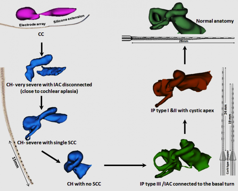
Malformed cochlear anatomy can present unique challenges, even for experienced surgeons. There is the is the initial challenge of identifying the malformation, then selecting an appropriate electrode for this unique cochlea, and then placing the electrode correctly in this challenging anatomy.
For example, in one malformation case, post-operative imaging showed the electrode array had entered the semicircular canal of the vestibular organ. Upon reimplantation, the electrode array entered the internal auditory canal (IAC), which is a further complication.
Electrode Array for Cochlear Malformation
Furthermore, choosing the appropriate electrode can be challenging in these cases. We know that individualized electrode selection is important for every cochlea, as one size does not fit all. This is especially important with inner ear malformations.
Let’s take this case as example. It looks like incomplete partition type I where you can expect to cover 360° of the available basal turn with an electrode. The surgeon chose a 24 mm long electrode hoping that the electrode will fit fully inside the cochlea.
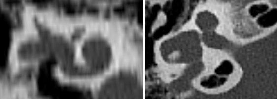
But it turned out that the basal 2 contacts did not go fully inside the cochlea. As a surgeon, you may not be super happy with the situation. We took the pre-operative image of this case and 3D segmented the cochlea and found that it was not that big to accommodate a 24 mm long electrode, but rather a 15 or a 19mm long electrode.
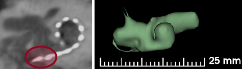
Post-operative CT indicates 2 electrode contacts are extracochlear. In this case, a FORM 19 or Compressed electrode array could have offered a better fit.
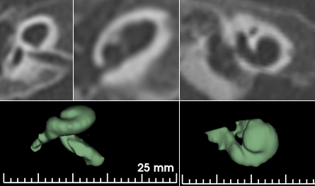
This is another case with malformation; this one is hypoplastic cochlea. We did the 3d segmentation and found that this is good enough to take an electrode array which is not longer than 20 mm. A FORM 19 was suggested and the surgeon successfully inserted fully inside the cochlea.
3D Printed Cochlear Models
3D segmentation of the cochlea from the clinical CT images is highly possible. We felt axial view is the best view for segmenting the cochlea. You just need to find the right grey scale threshold in capturing the inner ear and you get the 3D model of the cochlea. For educational purpose, we also 3D printed all the malformation types.
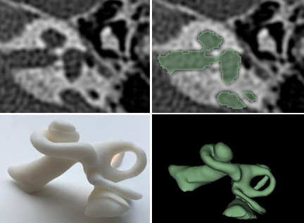
Creating a 3D printed model of a cochlea with enlarged vestibular aquaduct using CT images.
A 3D image adds more information than the 2D images; for example you can see how big the vestibular aqueduct is enlarged, and it gives the complete 360° view of the cochlea which could be of additional help to surgeons who have not seen many malformation cases in their career.