Join Clinicians Worldwide
Evidence-based insights to enhance hearing care—twice a month
Subscribe Now Evidence-based insights to enhance hearing care—twice a month
Subscribe NowMED-EL
Published Mar 07, 2019

We’ve always been scientists at heart. More than 40 years ago, our founders Ingeborg and Erwin Hochmair changed the world when they invented the modern microelectronic multichannel cochlear implant.
Their pioneering spirit and passion for discovery has always defined MED-EL. That’s why we devote so much of our resources into research & development. Our research initiatives are continually driving discovery and innovation in the field of hearing and beyond. The future holds so much potential, and we’re excited to be at the forefront of incredible breakthroughs and leaps in technology.
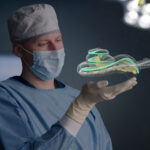
Today we’ve got a fascinating article from Daniel Sieber, Head of MED-EL’s Electrodes and Surgical Research & Development. Recently, Daniel and his research partners had their anatomical research published in Nature’s Scientific Data open access journal.
For the past four years, they have worked to create OpenEar—fantastically detailed 3D modeling of eight unique temporal bone specimens, including high-resolution modeling of:
Can’t See This Video?
Having problems viewing this video? Watch it on YouTube.
These new 3D models of the cochlea and related anatomy are completely free and open access. You can find the download links for the data and an open access 3D medical imaging viewer at the end of this article.
Let’s go over to Daniel to hear why the OpenEar project was so necessary, how there’s so much potential beyond surgical simulators, and why image-guided surgery & individualized cochlear implants are quickly becoming the standard of care worldwide.
My background is in engineering but in the last 11 years of working with MED-EL the scope has developed far beyond just technology and into a fruitful partnership with engineers, surgeons and healthcare professionals around the globe. My team is constantly pushing the limits of what can be achieved in cochlear implants—for example, we have developed intelligent solutions for surgery, individualized therapies, drug delivery systems, image guided, and robot-assisted surgery.
We also look at how we can support the best outcomes in cochlear implant surgery. One of the main challenges for cochlear implant surgeons is the amount of training and experience needed. This is because novice surgeons worldwide need to know the anatomy and surgery of the human temporal bone by heart before being able to start supervised surgery in patients. On the other hand, in many places it has become difficult to provide a sufficient number of anatomical specimens from donors for training. It can therefore be challenging for beginner surgeons to reach enough repetitions in a training environment to reach a level of automaticity in their surgery.
To help alleviate this problem, surgical simulators dedicated to surgery of the human ear have been developed by several groups worldwide during the last decade. Surgical simulators can be of great help in this situation because their digitized models of the ear never break or wear out. Like flight simulators in aviation, they provide an entirely hazard-free environment for training and at the same time allow for practically unlimited repetitions of procedures—self-directed and free of running cost.
MED-EL has for many years been in close touch with the researchers behind the Visible Ear Simulator system. Our high-tech temporal bone lab at headquarters in Innsbruck, Austria has the simulation integrated into a powerful virtual reality environment with haptic feedback devices and projection into the binoculars of latest generation digital operation microscopes.
Digital models of the ear also have great value beyond surgical simulation. Three-dimensional models are just much easier for our brain to understand, compared to looking at two-dimensional images which we are still looking at in medicine most of the time. Exploring anatomy in 3D just adds so much to the understanding of anatomy compared to looking at a textbook or CT scan.
When we think about the middle and inner ear for example, those are embedded in hard bone so they can not be easily seen in specimen. Two dimensional images found in textbooks on the other hand are often unprecise and entirely lack the information of where structures are in depth. Digital models allow to view and comprehend the anatomy by making the surrounding bone transparent and see structures in space and in relationship to each other. Furthermore, digital models created for surgical simulators can also be used in research related to image guided surgery, artificial intelligence and other applications.
However, for a long time the research community around surgical simulators has been confronted with a shortage in good quality digital models of the human ear. Most simulator systems relied on reconstructions from clinical CT scans. Such models lack a lot of details, for example, soft tissue detail and the entire lack of natural coloring of the models, as CT imaging is unable to capture color. This lack of colors makes the surgical simulation less realistic and more difficult as one can get lost without any texture references.
The Visible Ear Simulator is until today the only system which relies on a color model which had been created in 2002 by Prof. Mads Sølvsten Sørensen using cryosection and an extremely laborious process. So after more than 15 years the community was still left with just one single anatomy available in color.
I had been in touch with Mads and Peter Trier Mikkelsen, the software engineer behind Visible Ear Simulator since 2008 and somehow became part of the development during my spare time. We discussed many ways of how to improve the simulator, but one of the issues which we could not overcome was the lack of anatomical variation because of the limitation to just one individual anatomical specimen.
In 2012, I moved into a new position at MED-EL as Scientific Director of the Research Center Hannover coordinating many exciting research projects with Prof. Thomas Lenarz and his team. Suddenly I was allowed to spend many hours in the temporal bone lab and got in touch with the ingenious Peter Erfurt, who amazed me by constantly perfecting the artform of working with human tissues.
Finally, in 2014, the idea was born to try to create a new library of human temporal bone models using the epoxy embedding and grinding technique used in Hannover. The idea was to combine the advantages of CT imaging and micro-slicing of specimen to create a more efficient process for creation of new models.
CT is great because it can easily be segmented into different anatomical structures. Micro-slicing is great because it results in color images and shows non-bony structures such as the tympanic membrane which are not visible in CT. So, we performed CT and micro-slicing on the same bones and then performed a fusion of both images ending up with one dataset containing both imaging modalities.
This may sound straight forward, but it ended up in thousands of hours of work to develop the details—mostly from the spare time of people. Finally though we were able to provide colored models, and much faster than with the previous technique and thus yielding more anatomies.
Already at an early stage of the project we decided together with Dr. Ingeborg Hochmair and Prof. Thomas Lenarz to make the result of the project available open access/data. We were convinced it would be a waste of time and energy to create this kind of data and then let it dust in our files. Instead we published our process with Nature’s Scientific Data Open Access journal and the data on the Zenodo Open Data repository. The data can be used with many software packages capable of processing 3D images. However, we recommend using the free open source 3DSlicer software, which was also used in creation of the dataset.
We were able to collect data from eight different human temporal bones which are now in the process of being implemented into surgical simulators. Anatomical variation in the inner ear is huge and has large effect on the surgery, so with the eight new anatomies we are really just touching the tip of the iceberg of anatomical variation. But it already allows surgeons to train in a certain variation of anatomies and we hope this will improve their training.
This is essential, because when it comes to ear surgery, the smallest structures are those which are the most important for the hearing performance of patients. Keeping them unharmed is the big goal of modern-day soft surgery techniques. The entire inner ear is just the size of a pea, and some of the structures we were trying to display are only a few microns thick and would not show up on a CT scan alone.
Can’t See This Video?
Having problems viewing this video? Watch it on YouTube.
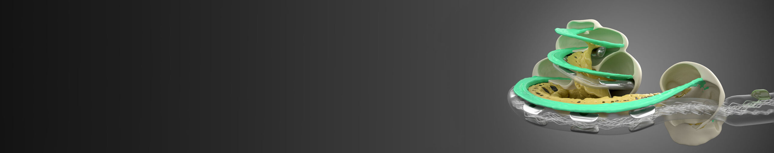
OpenEar 3D models allow you to explore 8 unique anatomical specimens. Segmentations of scala tympani, scala vestibuli, malleus, incus, stapes, facial nerve, chorda tympani, tympanic membrane, external auditory canal, sigmoid sinus and dura, carotid artery and bone are provided at a 125 μm isotropic voxel resolution as 3D NRRD images. You can view the models with any of the co-registered imaging modalities in the background. Additionally, triangulated models of those segments are provided with an optimized mesh quality using about 70 vertices/mm2 surface area in Polygon File Format (PLY) format. This can also easily be converted to the common STL file format using 3D Slicer, allowing for 3D printing of these anatomical models.
I’m absolutely excited about the quality of the images that resulted from the OpenEar project. I have to say that creating this dataset was hard work, at times using up our entire spare time. And of course, the realities of hundreds of hours of grinding specimen in the lab are not as glamourous as it seems when reading the publication. But I feel incredibly blessed and humbled being allowed to watch those thousands of slices of anatomical perfection unfold in front of me with every grinding step. I will be able to look at this data for years to come and find new aspects in them and I hope the research community will do so as well.
It was incredible to first-hand experience human anatomy on a microscopic level. The beauty and perfection of even the smallest structures in the human body are just breathtaking. It took about seven minutes to remove one layer from the anatomical specimen and prepare them for microscopy. Sometimes I could hardly find the patience to wait these seven minutes for the next layer to reveal itself and its wonders and would only be able to stop at 3 o’clock in the morning, just to be back at 9 to resume my work. I am truly grateful for this experience even though I have to admit I am also glad this time passed by, and I get more sleep again these days.
Today we are profiting from much better hardware and software when it comes to processing imaging data. Back in 2002, Mads performed manual segmentation on color images, which basically meant assigning each pixel in the image to an anatomical structure manually. Today we are using semi-automatic image segmentation techniques which rely on a registration of CT and color images. This is only possible because of improvements in registration algorithms and hardware improvements like e.g. GPU computing. I think we are now at the eve of artificial intelligence becoming much more usable for our research and so I think we are looking at an acceleration in our progress.
My belief is that within the next years, clinicians will be able to perform a CT scan and be presented with a three-dimensional view of the segmented relevant anatomical structures within minutes. This has particularly proven beneficial in cases of malformations of the anatomy, which can sometimes make it hard to understand the geometry in 2D images especially for less experienced surgeons.
This is so important for CI patients, because clinical data from various centers worldwide suggests that even today, we can maximize performance of cochlear implant recipients when choosing the optimal electrode and stimulation modality based on the candidate’s individual anatomy and pathology. Users show better hearing performance and more natural sound quality when these factors are taken into consideration.
It was an incredible ride to be allowed to lead this project, I am very grateful to the founders of MED-EL for allowing me to devote time and resources to the OpenEar. This clearly showed their dedication to understanding the human ear even better and moving the surgical field forward as a whole. Ingeborg and Erwin Hochmair are truly on a mission to overcome hearing loss as a barrier to communication and quality of life. They were the pioneers of structure preservation from the designer’s side and will be known also as pioneers of individualized cochlear implantation and image-guided surgery.
Once the surgeon can assess the 3D structure of the cochlea within minutes after taking a CT scan without manual process steps, today’s gold standard of individualized, anatomy and evidence-based treatment will become standard of care in every clinic worldwide and we are working hard every day to make this a reality as soon as possible.
These new 3D models of the cochlea and related anatomy are completely free and open access. OpenEar may be shared, used and adapted even for commercial use with the requirement of attributing to the original work as per a Creative Commons Attribution 4.0 License.
The original publication can be found here: The OpenEar library of 3D models of the human temporal bone based on computed tomography and micro-slicing [Article]
You can download all 8 models here: The OpenEar library of 3D models of the human temporal bone based on computed tomography and micro-slicing [Data]
The 3D anatomical models can be used with many software packages capable of processing 3D images. However, we recommend using the free open source 3DSlicer software [Viewer].
*Not all products, indications, and features shown are available in all areas. Please contact your local MED-EL representative for more information.
MED-EL
Was this article helpful?
Thanks for your feedback.
Sign up for newsletter below for more.
Thanks for your feedback.
Please leave your message below.
CTA Form Success Message
Send us a message
Field is required
John Doe
Field is required
name@mail.com
Field is required
What do you think?
The content on this website is for general informational purposes only and should not be taken as medical advice. Please contact your doctor or hearing specialist to learn what type of hearing solution is suitable for your specific needs. Not all products, features, or indications shown are approved in all countries.
MED-EL
Get the latest research and resources to help people with every kind of hearing loss. Subscribe to the MED-EL Professionals Blog now.
Registration was successful
We’re the world’s leading hearing implant company, on a mission to help people with hearing loss experience the joy of sound.
Find your local MED-EL team
The content on this website is for general informational purposes only and should not be taken as medical advice. Please contact your doctor or hearing specialist to learn what type of hearing solution is suitable for your specific needs. Not all products, features, or indications shown are approved in all countries.